

Kate Grieve, Scientific Director of the “Paris Eye Imaging” ocular imaging unit (www.pariseyeimaging.com), which she co-founded with Prof. Michel Paques and Prof. José Sahel at the CHNO Clinical Investigation Center of Quinze Vingts / Institut de la Vision, has just been awarded by the European Commission a 5-year “ERC Consolidator Grant”, which will enable her to pursue the development of optical imaging. Her “OPTORETINA” project aims to develop non-invasive optical measurements of retinal cell function in the living human eye. This development will help to significantly improve the diagnosis and monitoring of ophthalmological pathologies, as well as to evaluate in real time the results of innovative gene and cell therapies.
For patients with inherited retinal diseases, gene and cell therapies offer hope in preserving or restoring vision. Retinal imaging is crucial, first for the phenotyping of patients to determine which cells are degenerated and to design an appropriate therapeutic path, and then to monitor therapeutic success in patients who have been treated with these therapies. To date, the imaging tools used in the clinic do not provide sufficient resolution to visualize individual cells non-invasively, constituting a major obstacle to the development of these therapies.
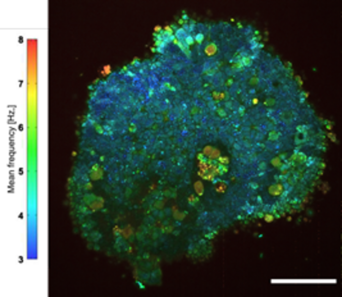
With her group, Kate Grieve aims to develop new tools for non-invasive cell imaging, such as full-field optical coherence tomography (FFOCT) and ophthalmoscopy with adaptive optics (AOO). Recently, she also devised a dynamic method of FFOCT using the movement of intracellular organelles, to detect metabolic contrast and thus indicate cellular activity. These new, non-invasive optical tools have the potential to provide, for the first time, subjective and objective measures of retinal function. In the "OPTORETINA" project, these tools will be adapted to stimulate the retina with visible light to allow testing of the visual function of patients with hereditary retinal diseases, including those treated in our clinical center with new gene and cell therapies, to verify that vision is preserved or restored successfully.
Contact
Kate Grieve PhD, Scientific director
Paris Eye Imaging
Clinical Investigation Center
Quinze Vingts National Ophthalmology Hospital
28 Rue de Charenton
75012 Paris
www.pariseyeimaging.com
e-mail: kgrieve[at]15-20.fr