vision-research.eu - The Gateway to European Vision Research
![]()

![]()
You are here: vision-research.eu » Vision Research » Vision in the European Focus » Vision in the European Focus - Details
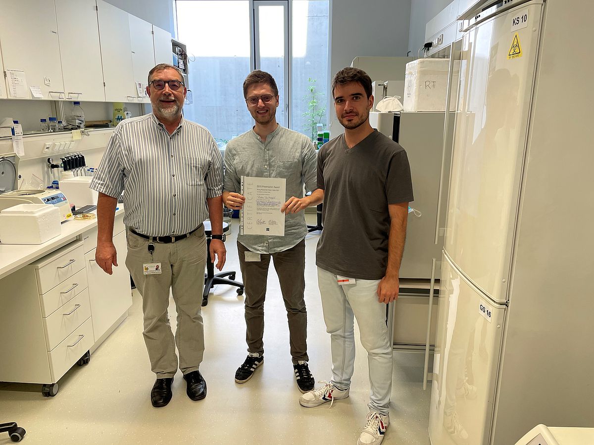
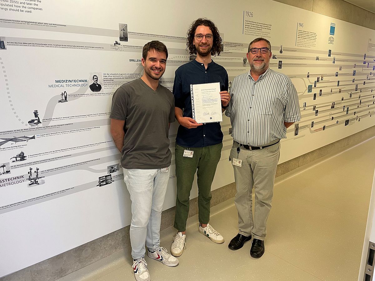
ZEISS Presentation Award @ Young Researcher Vision Camp 2021

This year the Young Researcher Vision Camp was performed as an on-line event. Therefore, the famous interactive poster session was transformed quickly in a “best talk” event with a broad spectrum of excellent contributions by the young researchers.
The award jury had an extremely difficult tasks to select the three best talks and recognize them with the ZEISS Presentation Award. Three awardees were chosen to reflect the high level of excellent contributions.
“There are only winners, and all young speakers did an excellent job”, said Thomas Wheeler-Schilling, CEO of the European Vision Institute, and organizer of the camp. Special thanks go to ZEISS Vision International for their generous support of the Young Researcher Vision Camp 2021 and the offer of the presentation award, which was given by Miguel Garcia Garcia, representing ZEISS, to the three awardees.
1st Prize goes to Gwen Musial
 |
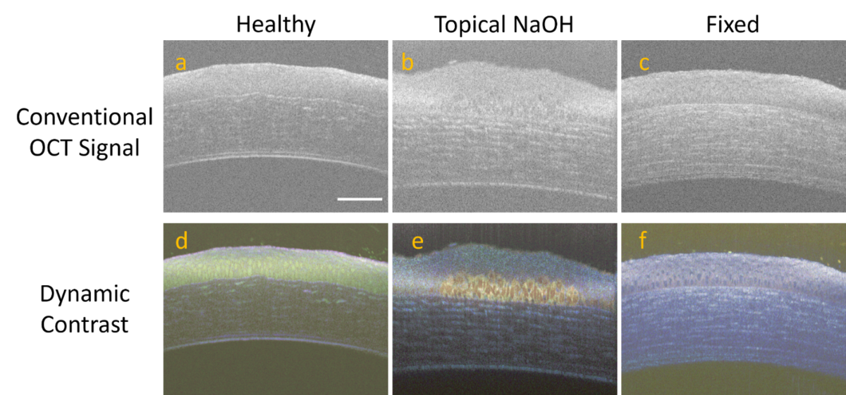
Dynamic contrast as a novel method of in-vitro tissue analysis
Gwen Musial, Hinnerk Schulz-Hildebrandt, Tabea Kohlfaerber, Uta Gehlsen, Gereon Huettmann, Philipp Steven
Background
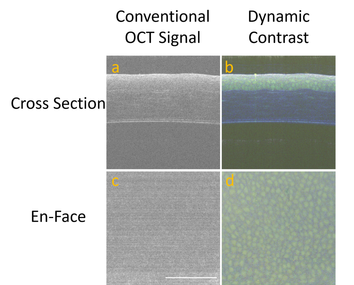
Optical coherence tomography (OCT) is a versatile imaging technique which is based on interferometry to detect backscattered light [1]. Using a broadband light source and a high numerical aperture objective, OCT imaging systems can be configured to have micron level resolution in both the axial and lateral plane [2], [3]. However, if there is a lack of scattering contrast in the desired imaging target due to only subtle or no changes in refractive index, it can still be difficult to image cellular structures. Dynamic OCT utilizes software that evaluates signal fluctuations due to ATP-dependent motion of subcellular structures to create images comparable to histological images [4]. While micro-OCT has been used to image cornea structures in vivo by several research groups [2], [5], [6], the combination of micro-OCT with the dynamic OCT software presents a unique opportunity to evaluate changes in cornea epithelium cell dynamics.
Methods
The custom-built Fourier domain system for microscopic OCT used in this study achieves a resolution of approximately 1 μm in all spatial dimensions [2]. Repeated B-Scans were acquired at a 100kHz A-Scan rate over a 0.5mm region with 512 A-scans per B-Scan. At each voxel, the temporal variations of the absolute value of the OCT signal were evaluated. The time series was Fourier transformed, and the integral amplitude was calculated in three frequency bands [7]. The pixels were color-coded in an RGB image, representing different time scales of motion activity. Eyes were taken from wild type black 6 (C57BL/6JRj) mice ages 12-20 weeks. Eyes were glued to a petri dish with tissue glue cornea facing up. Then the petri dishes were filled with warm cell culture media. While not being imaged, eyes were kept in an incubator set to 37˚C. To test eyes after an insult to the cornea, 10 µL of 0.4 M NaOH was applied to the eye to induce an alkali burn. To image eyes with no intracellular motion, healthy eyes were immersed in formaldehyde for fixation for one hour prior to imaging.
Results
Cornea epithelium cells were clearly visualized in dynamic contrast micro-OCT but were challenging to see in conventional OCT images (Fig 1). Application of NaOH and fixation in formaldehyde resulting in distinct changes to the dynamic contrast micro-OCT images (Fig 2).
Conclusion
This preliminary work shows promising early results for the use of dynamic contrast micro-OCT imaging as a novel method to evaluate corneal epithelium cells in-vitro. Further work will quantify the signal changes that arise from the application of NaOH to create an objective metric of cellular activation state.
 |
 |
2nd Prize goes to Pietro De Angeli
 |
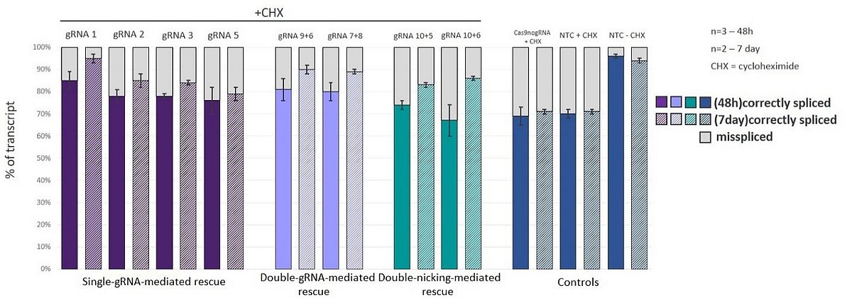
CRISPR/Cas9-based rescue of deep-intronic ABCA4 variant c.5197-557G>T in patient-derived cone precursor cells
Stargardt disease is an autosomal recessive inherited retinal disorder caused by biallelic mutations in ABCA4. Deep-intronic variants (DIV), resulting in aberrant mRNA splicing, have been associated with an increasing number of cases. Upon splicing, DIV c.5197-557G>T causes a 188-bp pseudoexon insertion in the mature mRNA which is degraded via nonsense-mediated decay. Here, we aim to investigate, assess and compare three different Cas9-based strategies to rescue this splicing defect in patient-derived cone precursor cells (CPCs) by genome editing.
Three strategies employing Streptococcus pyogenes Cas9 (SpCas9) were designed. The gDNA cut efficiency was assessed in either HEK293T cells or patient-derived fibroblasts. The ability to rescue the correct splicing was then studied with minigene assays in HEK293T cells. Patient-derived human induced pluripotent stem cells were differentiated into 2-dimensional CPC cultures with a 30-day differentiation protocol. Successful Cas9 strategies were transfected in CPCs via electroporation. Fluorescence-activated cell analysis was used to measure the transfection efficiency, and the mRNA splicing rescue was quantified with fragment analysis.
In short, splicing correction of c.5197-557G>T was successfully achieved in CPCs. Heterozygous ABCA4 c.5197-557G>T-patient-derived CPCs showed a background expression of the wild type allele of 71%±1 of total ABCA4 transcript. Upon Cas9 delivery, the total expression of the wild type allele increased to 95%±2 (0.1 mg/ml cycloheximide, n=2). These results show for the first time the possibility of targeting DIVs in patient-derived CPCs with CRISPR/Cas9 strategies. Ultimately, our work can pave the way to ex-vivo editing of patient-derived CPCs prior to autologous transplantation and it can offer a powerful platform for the study of personalized Cas9-based therapies targeting photoreceptor cells.
 |
 |
3rd Prize goes to Yannick Sauer
 |
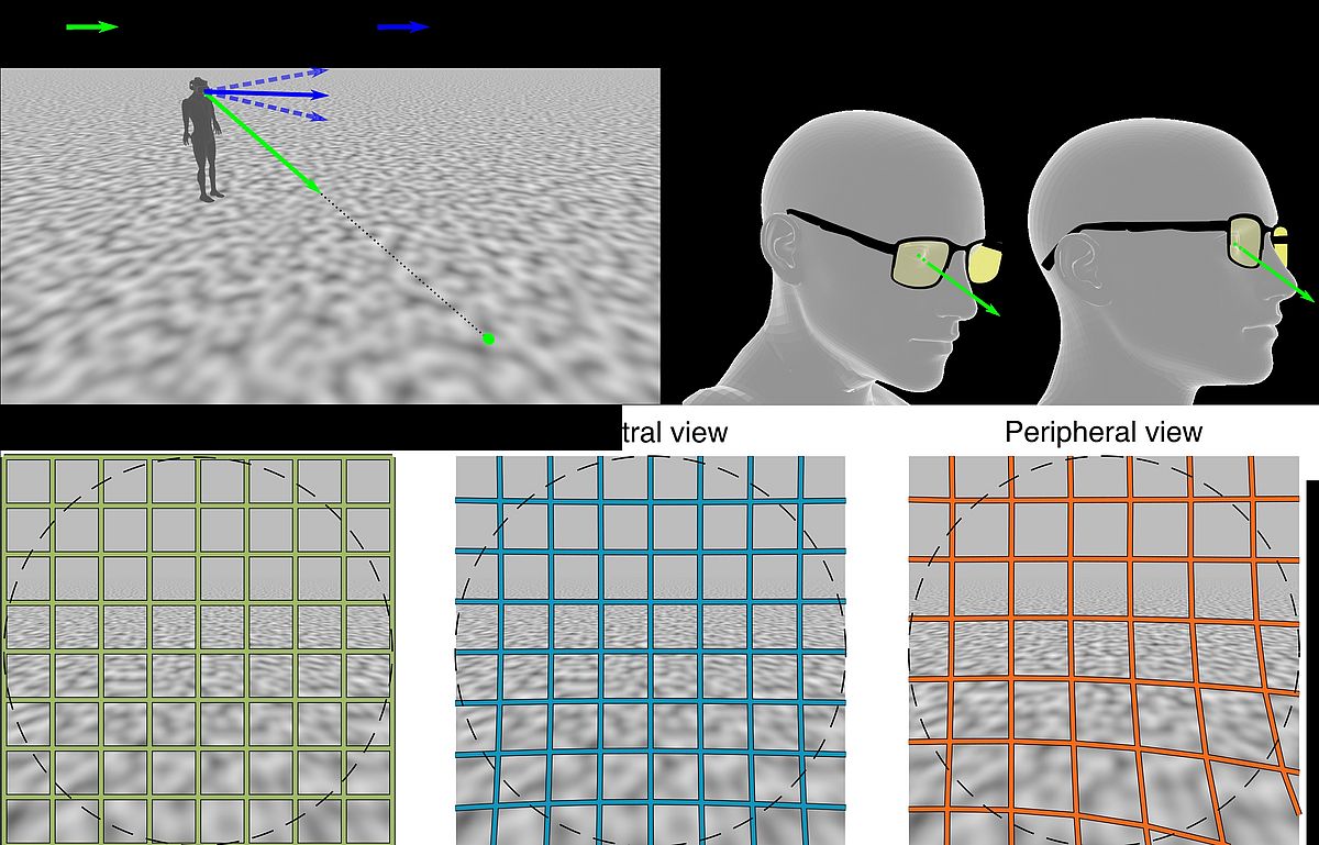
Studying visual perception under the influence of distortions in a VR-based optical simulation
With increasing age the accommodative capability of the human eye decreases, it gets harder to focus close objects. Progressive addition lenses (PALs) are a common treatment method. This special kind of spectacle lens has di_erent zones of di_erent optical power for far and near vision. Between the zones there is an progressive increase in optical power. As a side e_ect, this will always lead to unwanted optical aberrations causing blur and distortions in the visual _eld. PAL wearers often report about distortion related discomfort and the perception of unnatural motion.
To study the inuence of distortions on visual perception we developed a virtual reality (VR) based simulation. The rendered image is transformed to present a distorted _eld of view equal to the distortions perceived by PAL wearers. The advantage of simulating distortions in VR is the possibility of reproducible conditions equal for every subject and easy access to behavioural data of eye- and head-movements.
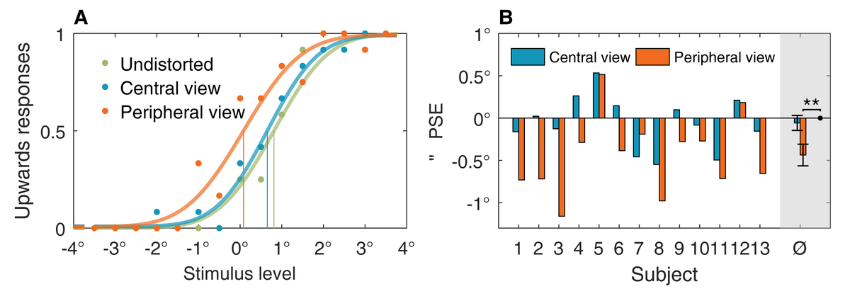
One possible inuence of distortions on perception occurs during self-motion. Humans are able to determine their motion direction from visual input. Distortions change this input in a complex way, making it hard to predict the inuence of distortions on self-motion perception. We studied a typical self-motion scenario of walking on a ground plane. In this case gaze is typically on the ground a few meters ahead of us. To _xate a point on the ground, multiple combinations of head- and eye-orientation are possible: one could keep the head straight and move the eyes down or bow the head while eyes are straight relative to the head. The visual input in both conditions is the same, since we see the same part of the moving oor. For PAL wearers though, the two conditions lead to a di_erent perception of distortions. When gaze is through the center of the lens, only the periphery of the visual _eld is distorted. For gaze through the periphery of the lens more distortions are visible. We recreated this scenario in VR by moving subjects virtually along a ground plane while simulating distortions either for the central or the peripheral view through a PAL. Perception of vertical motion direction was tested by combining the forward motion with a small angle upwards or downwards. In repeated trials, subjects answered if the motion they perceived was upwards or downwards. The answers depending on stimulus level can be _tted to _nd the point perceived as straight in each distortion condition. Results show that in average in the peripheral view condition a downwards motion was perceived as straight. This means motion is perceived more upwards than it actually is. In the central view condition there was no signi_cant e_ect indicating that distortions only in the periphery have less inuence on self-motion perception.
In conclusion, this experiment showed that distortions can inuence self-motion perception and the perceived direction depends on which part of the lens one uses. This conicting sensory input might be one reason for the discomfort or even nausea some PAL wearers su_er from.
 |
 |
 |
 |